
おかげさまで開設25周年oscestop.education 創業祭
oscestop.education


 お店で受け取る
(送料無料)
 配送する
配送する
納期目安:
01月25日頃のお届け予定です。
決済方法が、クレジット、代金引換の場合に限ります。その他の決済方法の場合はこちらをご確認ください。
※土・日・祝日の注文の場合や在庫状況によって、商品のお届けにお時間をいただく場合がございます。
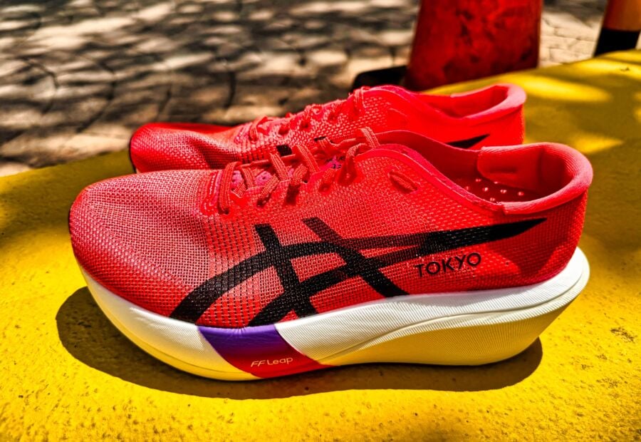
ASICS METASPEED EDGE TOKYO 24.5 Amazon.com | ASICS Unisex METASPEED Edge Tokyo Running Shoesの詳細情報
Amazon.com | ASICS Unisex METASPEED Edge Tokyo Running Shoesスパイク・シューズ ASICS SPEED EDGE TOKYO 24.5 Cut in half
ほぼ新品です。
走行距離は8kmです。
いきなりの値下げ交渉はご遠慮ください。
#メタスピード
#メタスピードTOKYO
#エッジ
#ランニングシューズ
#マラソン
#トレーニング
#アシックス
商品の情報
| カテゴリー | スポーツ > 陸上競技 > スパイク・シューズ |
|---|---|
| ブランド | asics |
| 商品の状態 | 未使用に近い,数回使用し、あまり使用感がない |


ベストセラーランキングです

![Spit: Squeegee Punks in Traffic [DVD]](https://img.fril.jp/img/758158201/l/2560469007.jpg?1744998297)
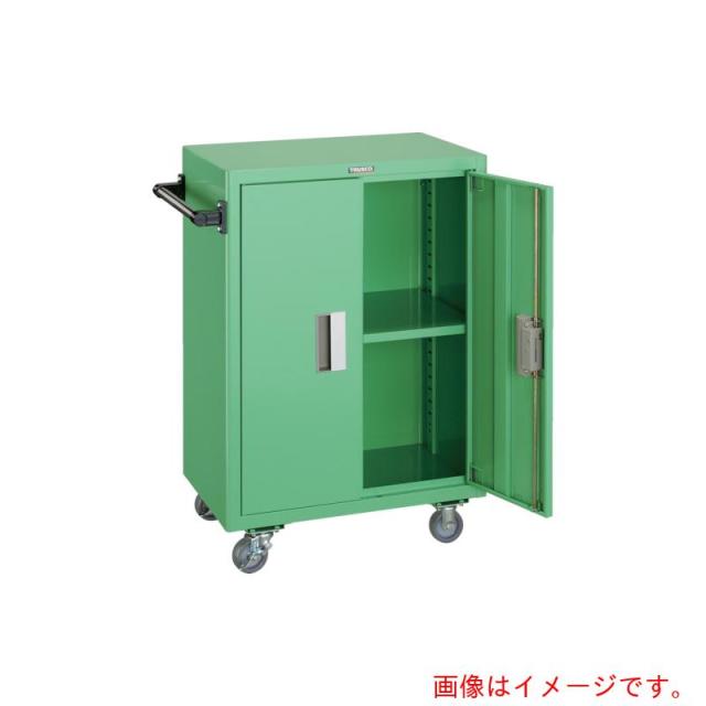
![岩田製作所 【個人宅不可】 IWATA ラバーエッジトリム 133M TRE08-L133 [806-440715]](https://tshop.r10s.jp/daishinshop/cabinet/item/806-67/806-440715.jpg)

この商品を見た人はこんな商品も見ています
-
 マイストア在庫: 15税込17,269円
 -
 マイストア在庫: 15税込6,621円
 -
 マイストア在庫: 15税込20,318円
 -
 マイストア在庫: 15税込6,831円
 -
 マイストア在庫: 15税込7,289円
近くの売り場の商品


カスタマーレビュー
オススメ度 4.6点
現在、77件のレビューが投稿されています。