Share your insights

Help us by sharing what content you've recieved in your exams


Cranial nerves examination

NB: you may be asked to examine only the visual cranial nerves (CN 2, 3, 4 and 6) or the bulbar cranial nerves (CN 9, 10 and 12).

Introduction

  • Wash hands
  • Introduce self
  • Ask Patient’s name, DOB and what they like to be called
  • Explain examination and obtain consent

General inspection

  • Look at the patient in general from the end of the bed
    • Posture
    • Habitus
    • Other signs of neurological conditions
  • And look around the bed for signs such as:
    • Mobility aids
    • NBM signs
    • Fluid thickener
    • Glasses
    • Hearing aids

1. Olfactory

  • Test each nostril with smelling salts if available
  • If not, ask patient if they have noticed any change in smell

2. Optic

Start with inspection (visual aids; pupil size and symmetry; screening test – ask if they can see your whole face clearly). Then AFRO (3 tests for each):

Acuity

Ask the patient to cover one eye with their palm and test each eye in turn

  • Distant vision (visual acuity): test with Snellen chart (the result is recorded as distance/smallest font size read, e.g. 6/9)

    • If the patient wears glasses, test with glasses on (corrected visual acuity)
    • A standard Snellen chart is read from 6 metres away but there are smaller versions which may be used at closer distances (e.g. 1 or 3 metres) – adjust the final acuity to ‘1/…’ or ‘3/…’ respectively
    • If the patient gets more than two letters wrong, the previous line should be recorded as their acuity. If they get two letters wrong, record acuity as the font size of this line but note ‘-2’ in brackets, e.g. 6/9 (-2); and if they get one letter wrong, note ‘-1’ e.g. 6/9 (-1).

  • Near vision: read a line of a letter/magazine
  • Colour vision: ‘I would also like to test colour vision using Ishihara plates.’

Fields

Sit the patient 1 metre directly in front of you with both your eyes at the same level

  • Visual inattention: while the patient keeps both eyes open and focussed on you, hold out your hands in each of their outer visual fields. Ask them to point at the hand(s) which you are opening and closing. (Inattention to one side, i.e. identification of only one moving hand when both are moving = contralateral parietal lesion.)
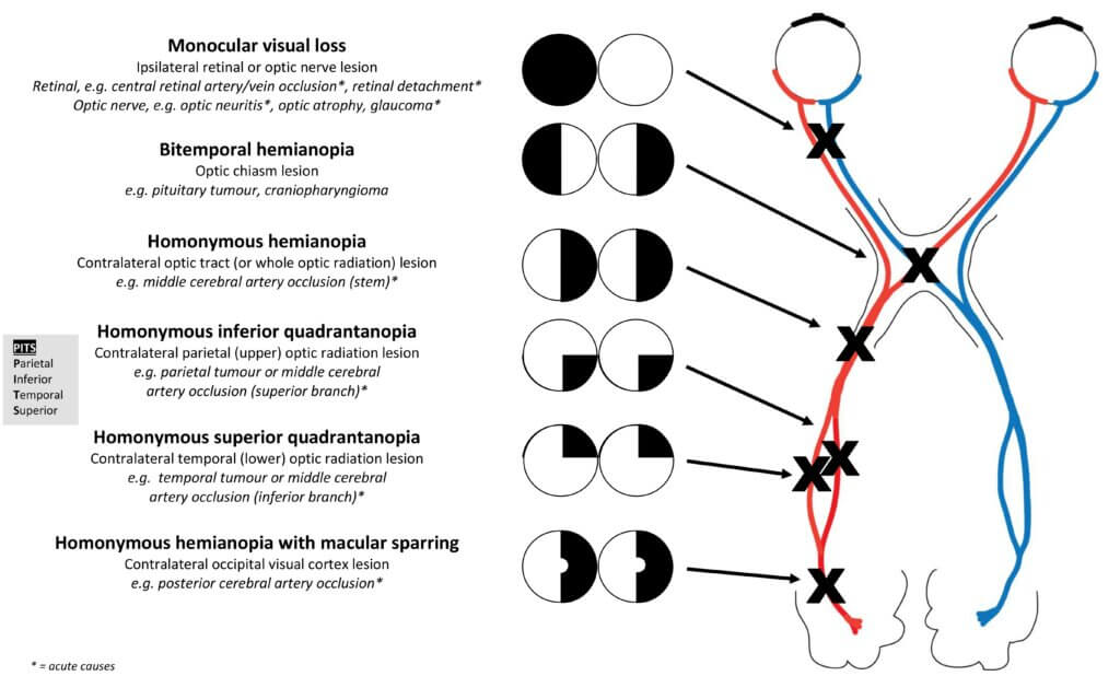
  • Visual fields: ask the patient to cover one eye with their palm and close your eye on the same side (without using your palm if you can). Ask them to stay focussed on your open eye. Select a white visual fields pin and bring it in from the periphery, keeping it at mid-distance between you and the patient. Ask them to tell you when they can see it. Move in a diagonal direction into each of the four quadrants. Test both eyes individually, comparing their fields with yours.

    • Mononuclear field loss = intra-ocular pathology or ipsilateral optic nerve lesion
    • Bitemporal hemianopia = optic chiasm compression
    • Left/right homonymous hemianopia = contralateral optic tract/radiation lesion, or occipital cortex if macular sparing is present

  • Blind spots (offer to test): while the patient keeps both eyes open and focussed on you, hold a red pin mid-distance between you. Check they can see it as red in the middle (central scotoma = optic nerve lesion). Now move the pin horizontally towards the periphery in each direction and to tell you when it disappears. Map each of their blind spots against your own (large blind spot = papilloedema).

Reflexes

  • Accommodation: ask the patient to focus on a distant object, then hold your finger close to their face and ask them to focus on it. Pupils should constrict and eyes should converge.
  • Direct and consensual papillary reflexes: in a dimmed room, ask the patient to hold an open hand between their eyes and focus on a distant point in the room. Shine the light at each pupil in turn from about 45°. Observe for direct and consensual papillary constriction.

    • Afferent defect (i.e. pupils are symmetrical but when light is shone in the affected eye, neither pupil constricts) = CN2 (optic nerve) lesion
    • Efferent defect (affected pupil is persistently dilated, whilst other is reactive to light being shone in either eye) = CN3 lesion

  • Swinging light test: swing the light between the two eyes – the pupil size should stay the same regardless of which eye the light is shone in. If pupils become more dilated when the light is shone in one eye, then that eye is less sensitive to light and, hence, there is a relative afferent pupillary defect in that eye (partial optic nerve lesion on that side).

Ophthalmoscopy

‘I would also like to perform ophthalmoscopy to visualise the optic disc.’ See how to here.

Visual field defects

3,4,6. Oculomotor, trochlear, abducens

Ask if the patient has any double vision and to tell you if they experience any during the test. First inspect for strabismus and ptosis (partial = Horner’s syndrome; complete = CN3 lesion).

H-test

  • Ask patient to keep their head still (you may need to hold a finger on their forehead) and, with both eyes open, to follow your finger
  • Make an ‘H’ shape
  • Pause when they are looking laterally (nystagmus = cerebellar pathology)
  • If there is complex ophthalmoplegia, ask them to look straight up while counting down from 20 (fatigability suggests myasthenia gravis)

Saccades test

  • Ask the patient to look back and forth between two widely spaced targets, e.g. your index finder on one hand and thumb on the other, while keeping their head still
  • Test horizontally and vertically
  • Check for conjugate eye movements and target accuracy
  • Delay, inaccuracy, or slow or disconjugate movements suggest a central nervous system pathology rather than a peripheral vestibular pathology

Extraocular muscle lesions

CN3 supplies all extra-ocular muscles except Superior Oblique (CN4) and Lateral Rectus (CN6) – SO4LR6

If the eye cannot move laterally: there is a CN6 lesion

If the eye cannot move inferiorly when facing medially: there is a CN4 lesion

If the majority of the eye’s movements are impaired and the eye rests in a ‘down and out’ position: there is a CN3 lesion

If there are dramatically abnormal eye movements which do not fit with a single nerve lesion: there is ‘complex ophthalmoplegia’ (Graves/ mitochondrial/myasthenia/brainstem lesion)

5. Trigeminal

  • Inspect: temporalis/masseter muscle wasting
  • Sensory: ask the patient if they have any areas of pins and needles or numbness. With the patient’s eyes closed, use a cotton wool ball ± a neurological pin to test sensation over the ophthalmic, maxillary and mandibular distributions of the trigeminal nerve. Ask the patient to tell you when they feel it and if it feels the same on each side. 
  • Motor: ask the patient to clench their jaw and feel the bulk of the temporalis and masseter muscles. Ask them to open the jaw against resistance.
  • Others: ‘I would also consider testing the corneal reflex (afferent = CN 5; efferent = CN 7) and jaw jerk (afferent and efferent = CN 5).’

7. Facial

  • Inspect: forehead wrinkles, nasolabial folds, angles of mouth for facial asymmetry
  • Motor 
    • Raise eyebrows
    • Scrunch up eyes (try to prise each open in turn with your thumbs)
    • Purse lips together (try to prise each open in turn with your thumbs)
    • Show teeth
    • Puff out cheeks (try to push air out)
  • Others: ‘I would also consider testing taste on the anterior two-thirds of the tongue.’

UMN and LMN facial nerve lesions

  • The forehead is spared in UMN lesions because the nucleus controlling the upper part of the face has bilateral UMN innervation.
  • In a LMN lesion (e.g. Bell’s palsy), the whole side of the face is affected.

8. Vestibulocochlear

  • Crude hearing test: ask patient to occlude one ear and whisper a number into the other. Start at a distance and move towards the ear, asking them to tell you the number when they hear it. Repeat on the other side.

Weber’s test

  • Use a 512Hz tuning fork
  • Twang the prongs and place the round base of the fork on the patient’s forehead between their eyes
  • Ask them if one side is louder than the other
  • If one side is louder, either that side has a conductive deficit, or the contralateral side has a sensorineural deficit – Rinne’s test can then confirm which

Rinne’s test

  • Use a 512Hz tuning fork
  • Twang the prongs and place the round base of the fork on the patient’s mastoid process. Ask them to tell you when the sound stops
  • Then, place the prongs near the patient’s ear and ask if they can hear it again
  • Air conduction should be louder than bone conduction
  • If they cannot hear it again, there is a conductive deficit in that ear

  • Others: ‘I would also consider performing vertigo tests such as walking on the spot or vestibulo-ocular reflex testing.’

9,10. Glossopharyngeal, vagus

  • Inspect: ask patient to open mouth and say ‘ahhh’ while you use a pen torch to inspect palate symmetry (CN 9) and uvula deviation (CN 10) – uvula deviates away from side of lesion
  • Motor: assess speech, cough and swallow 
  • Others: ‘I would also consider testing the gag reflex (afferent = CN 9; efferent = CN 10) and taste on the posterior third of the tongue (CN 9).’

11. Accessory

  • Inspect: sternocleidomastoid/trapezius muscle wasting
  • Motor: ask the patient to turn their head to each side against resistance (tests contralateral sternocleidomastoid muscle), and shrug their shoulders against resistance (tests trapezius)

12. Hypoglossal

  • Inspect: tongue while relaxed in mouth for muscle wasting and fasciculations (LMN lesion, i.e. bulbar palsy)
  • Motor: stick out tongue (deviates to side of lesion – Tongue = Towards), move from side to side and test power by resisting tongue pressed into cheek

To complete

  • Thank patient 
  • ‘To complete my examination, I would perform full upper and lower limb neurological examinations.’
  • Summarise and suggest further investigations you would consider after a full history

Why don’t you test your knowledge?

What is the difference between a medical and surgical third nerve palsy? Name some causes of each.

Oops! This section is restricted to members. Click here to signup!

Discuss how you would clinically distinguish between an LMN facial nerve lesion and a UMN facial nerve lesion, and comment on why they present differently.

Oops! This section is restricted to members. Click here to signup!

Please list three causes of a unilateral facial nerve lesion, and two causes of bilateral facial nerve lesions.

Oops! This section is restricted to members. Click here to signup!

Now try out the skills you’ve learned on some real stations!

  1. Cranial nerve exam
  2. Bells palsy
  3. CN6 palsy
  4. Horner’s syndrome
  5. Bulbar cranial nerves exam
  6. There’s more here

No comments yet 😉

Leave a Reply